A vitreous hemorrhage may have different causes. In many cases, medical treatment is only possible to a limited extent. However, bleeding often regresses on its own.
What is vitreous hemorrhage?
If there is a vitreous hemorrhage, blood penetrates into the so-called vitreous space of the human eye. The vitreous body takes up approx. 80% of the available space in the human eyeball and is filled with a clear vitreous body fluid.
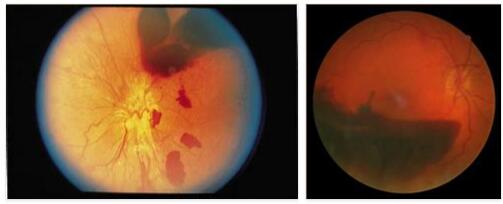
Vitreous hemorrhage can cause the vitreous humor to become cloudy. In those affected, this is often associated with impaired vision. Vitreous hemorrhage can take on different degrees: A slight vitreous hemorrhage manifests itself, for example, in the fact that there are few dark spots in the affected person’s field of vision.
If vitreous hemorrhage is very pronounced, it can restrict vision so severely that the patient can, for example, only distinguish between light and dark.
Causes
The most common cause of vitreous hemorrhage is what is known as diabetic retinopathy. This is a disease of the retina, the development of which is favored by an existing diabetes disease (sugar disease).
In young people, vitreous hemorrhage is also often caused by external injuries to the eye. Other factors that can cause bleeding into the vitreous are, for example, the presence of high blood pressure or cerebral haemorrhage. Vitreous hemorrhage can also be caused by a detachment of the retina, in the context of which it comes to bleeding of the retinal vessels.
Bleeding into the vitreous humor can also be a possible consequence of malignant neoplasms (such as vascular tumors) on the retina; a corresponding vascular tumor can lead to bleeding in the eye. Last but not least, various underlying diseases such as leukemia can ultimately promote vitreous hemorrhage.
Symptoms, ailments & signs
Vitreous hemorrhage symptoms depend on the extent of the vitreous hemorrhage. First of all, there are changes in image perception, which are expressed in dark appearing opacities. These localized opacities are caused by blood pockets under the vitreous humor. Patients describe them as black flakes, cobwebs, or particles in suspension.
Furthermore, those affected can perceive points, moving shadows or even flashes of light. The flashes of light already indicate a vitreous detachment. These soot-rain-like flakes that appear suddenly appear especially in the morning after getting up, as the blood moves back and forth violently under these conditions.
Sometimes sometimes occur even visual field defects, where individual parts of the overall perceived visual field appear blinded by blood retention. The field of vision is also discolored red from the blood. Overall, however, the vitreous hemorrhage does not cause any pain. If the bleeding is weak, the symptoms are limited to the symptoms described above.
Reductions in visual acuity then do not have to occur. However, if the bleeding is heavier, reversible vision deterioration may also occur. Reversible visual impairment means a temporary reduction in vision. Even with a blood volume of ten microliters, the visual acuity can be so severe that the patient can only see hand movements instead of sharp images.
In severe cases, reversible blindness is often observed. Vitreous hemorrhages can heal on their own. In the case of severe or recurring bleeding, the blood can no longer be reabsorbed, which can lead to tears in the retina and even to its detachment.
Diagnosis & course
A suspected diagnosis of vitreous hemorrhage by the ophthalmologist may initially be based on the patient’s complaints. In order to confirm such a diagnosis, an ophthalmological examination can be carried out with a so-called slit lamp or a slit lamp microscope, for example.
Here, a slit-shaped light beam is directed onto the patient’s eye in order to be able to detect a vitreous hemorrhage. If this examination method cannot deliver clear results, it is possible in a further step to detect vitreous hemorrhage with the help of ultrasound methods.
The course of a vitreous hemorrhage differs depending on the patient and the severity of the bleeding. In principle, it is possible for bleeding to subside on its own. It does this by the body reabsorbing the blood that has entered the vitreous humor. Such a process can take several months and beyond. In severe cases, vitreous hemorrhage can lead to spontaneous blindness in the affected person.
Complications
Vitreous hemorrhage is usually diagnosed late, which means that treatment is delayed. The symptoms usually only show up as the disease progresses, whereby the patient’s field of vision is restricted. This leads to black spots, which can limit the everyday life of the person affected. The eyeball can also turn red.
However, the restrictions in the field of vision often lead to psychological complaints and stress. Headaches and dizziness can also occur, which lead to a reduced quality of life. In the further course of the disease, the eyesight is reduced more and more, so that in the worst case, it can lead to a complete loss of vision.
This blindness is not reversible and can no longer be treated. If the vitreous hemorrhage is treated early, no particular complications will arise. The treatment usually takes place surgically and stops the bleeding.
However, this does not rule out the possibility that the bleeding will recur in the patient. Vitreous hemorrhage usually does not change life expectancy. Furthermore, cataracts can develop after the treatment.
When should you go to the doctor?
Since the vitreous hemorrhage is a serious complaint, it must always be examined and treated by a doctor. Early treatment can prevent further complications and eye discomfort. You should usually see a doctor if there is bleeding in the eye. In most cases, this bleeding is clearly visible so that other people can also alert the patient to the vitreous hemorrhage.
Most of the time there is no pain. Furthermore, the perception of the colors can change so that the field of vision looks reddish. A total loss of the visual field can also indicate a vitreous hemorrhage and must always be examined. There are also visual problems or even blindness.
The first point of contact in the event of a vitreous hemorrhage can be an ophthalmologist. If the complaint occurs after an accident, an emergency doctor can be called or the hospital can be visited directly. In most cases, this bleeding can be treated well so that there are no further complications.
Treatment & Therapy
Which therapy is used in individual cases to treat vitreous hemorrhage initially depends on the cause of the bleeding. If the bleeding is a symptom of an underlying disease, the consistent treatment of this underlying disease is one of the therapeutic goals.
If the ophthalmologist has found that vitreous hemorrhage is not the result of retinal damage (or that the retina is intact), doctors often advise first to wait for the blood that has entered the vitreous to break down naturally. One of the reasons for this is that the options for drug treatment of vitreous hemorrhage are very limited. One of the risks of not receiving therapy is that bleeding may continue.
In severe cases, vitreous hemorrhage can be treated surgically. Such an intervention can, however, promote a postoperative occurrence of retinal detachment or eye diseases such as cataracts. During a surgical procedure, the vitreous humor affected by the vitreous hemorrhage is usually removed. The vitreous humor is replaced by a liquid that can be based on a saline solution, for example.
Outlook & forecast
The prognosis for vitreous hemorrhage depends on the stage of the disease and the underlying disorder. In addition, spontaneous healing is documented for many patients, so that natural relief and subsequent freedom from symptoms can be achieved at any time, even without therapy.
The difficulty of the disorder lies in the early diagnosis and treatment options for the disease. The further the vitreous hemorrhage has progressed, the more unfavorable the future course of the disease is classified. Without medical treatment, a large number of patients gradually develop progressive loss of vision until they ultimately become completely blind.
This condition is irreversible and causes psychological problems or secondary diseases in most patients. If the retina is damaged, the prognosis is also worsened. Surgical intervention is necessary, which is associated with risks and side effects.
If the causal triggers of the vitreous hemorrhage can be treated well and the blood that has formed can drain away, the prospect of subsequent freedom from symptoms is good. This applies in particular to patients who have no severe visual impairment and who do not have any other eye diseases. Vitreous hemorrhage can occur again over the course of one’s life. The prognosis remains unchanged if the symptoms recur.
Prevention
Vitreous hemorrhage can only be prevented to a limited extent. As a rule, however, regular ophthalmological check-ups are important. This is because various diseases that can lead to bleeding in their course must be recognized and treated at an early stage. For example, a beginning retinal detachment diagnosed in good time can be stopped and thus vitreous hemorrhage can be prevented.
Aftercare
Vitreous hemorrhage follow-up consists of two parts. The eye in which the intravitreal bleeding took place must be checked again at least once, but usually several times, in order to rule out further bleeding and to observe the healing. If necessary – if medication was used – additional funds are prescribed. In addition, the eye should be checked for possible consequential damage. An ophthalmologist has the necessary equipment to also check the inside of the eye properly.
More importantly, however, is understanding the cause of the bleeding. While this is easy to do in the case of accidents and the application of force to the face, spontaneous vitreous hemorrhage is a reason for further investigation. For example, various diseases can be considered as the cause. These include, for example, unknown diabetes, retinal detachment or tumors.
The usual follow-up measures after surgical interventions on the eye are also necessary. This is especially true if vessels have been treated surgically or cracks in the eye have been treated. Wounds or bleeding in the eye take a long time to heal, which is why there should be several follow-up examinations. If necessary, agents can be prescribed to accelerate the breakdown of blood in the vitreous humor.
You can do that yourself
Patients with a vitreous hemorrhage are able to do self-help through various measures and thereby both alleviate the acute symptoms and positively influence the long-term course of the disease. It is fundamentally important to always follow the advice of the treating doctor.
In order to support the initial treatment with the aim of hemostasis, the patient maintains an upright, calm position. The head is therefore above the trunk and extremities so that the amount of blood collects in the lower area of the eye. During the illness, physical activity should be reduced so as not to worsen the course of the illness. The same applies to sports, especially strength training and competitive sports. Any sporting activity must be coordinated with the specialist. This increases the chance that the bleeding will recede more quickly and that the patient will regain his usual quality of life.
If surgical treatment of the vitreous hemorrhage is necessary, the affected person supports the chances of success of the procedure by adopting a healthy lifestyle. A strengthened immune system and consideration for the special hygienic requirements before and after the operation in the area of the eye promote healing. During the entire illness, the eyes must be spared and prevented from overexertion, for example by not exposing the patient to extreme weather conditions and by minimizing the use of screens.